HOME  | COURSES |  ALUMNI  | RESEARCH ACADEMICS  |  ABOUT US  |


Osteopathy in the Cranial Field, Osteo Craneo Field
Osteopathic Cranial Approach to Treatment
Sutherland Techniques on Craniosacral Therapy
Lato Sensu Ad Modum Sutherland
Scientific Study Hours: 150 Hours
Clinical Training Hours: 50 Lessons (à 45 min.) during 7 days


About the Course

This Course is the Foundation of Cranial Osteopathy

The course consists of a well-balanced mixture of short lectures followed by practical sessions.

The course will explore anatomy and theories of causation of the primary respiratory mechanism, the physiology of the Primary Respiratory Mechanism as well as contraindications and a protocol for treatment of the vault, diaphragms and sacrum.

Description

Lato Senso course comprising academic lectures, practical sessions, scientific study course work, and clinical literature review plus directed & guided home-study. The Course is based on the Original Teachings of Dr Andrew Taylor Still, DO.

Aims

Development of the knowledge & transferable skills required to begin applying the Philosophy, Principles & Concepts of Osteopathy in the Cranial Field in practice; and to stimulate further future academic and practical interest in studies relating to the Anatomical and Physiological functioning of the Physical, Mental and Spiritual bodies (how to same interact one with each other) and its relations to the Creator.

Please Note

The Course serves no intention to qualify the student has an Osteopathic doctor.
The Course is a Foundation Course on Osteopathy in the Cranial Field, Methods and Techniques.
School students who successful complete the course are trained to treat certain common conditions in a safe manner, in which Craniosacral Treatment is of benefit.

Assessment Criteria

Fulfilment of the learning outcomes relating to each module; and meeting the attendance expectation of 100%. Students who for any given reason who miss the certain class will have to take those missed classes before a Diploma is awarded.

Please note: Those students who miss classes and do not take at a later date those missed classes will be awarded a Certification of  Attendance to the Course stating the total of Hours attendance and which modules where attended.

Assessment Methods

Written course work, review presentation, case-history presentation, practical examination by viva.

Bibliography

Set texts & references in relation to each module; guided literature review.

Pre-Course Reading Required:

1. Craniosacral Therapy: What It Is, How It Works, John E. Upledger

2. Your Inner Physician and You: Craniosacral Therapy, John E. Upledger

3.
The Cranial Bowl, William G. Sutherland (if difficult to obtain or out of print, contact the school for a - not for sale - reading copy)

4. Course Manual.

Course Material

Course Materials in respect to the Teachings of Osteopathy in the Cranial Field, this are given free of charge, which are a fundamental part of the Teaching Materials.
Video teaching aids materials are provided for private use only and self study.

Prerequisite Qualifications

Minimum requisites: Students applying to this Course need to have Anatomy, Physiology and Pathology Course, either as a Course which was taken specific for Anatomy, Physiology and Pathology or part of another course, in which AP&P is given.

Please be aware that the minimum amount of Anatomy, Physiology and Pathology classroom hours should be no less than 150 hours.
Anatomy, Physiology and Pathology can be taken with Volksmed School or with any other school.

Diploma on Completion of Course

Diploma in Osteopathy in the Cranial Field, Foundation Course Theory & Practice

Students per Course

1 to 8 maximum (This course is very technical thus its not suitable for larger numbers in clinical training)

Course Fees:  £1,200
Price includes: Accommodation, Breakfast, Lunch & Dinner & Accommodation

Change of Mind & Refunds: Our courses are given to the best standards and are second to none.
But each person is different and we understand that things may change.

Our Guarantee to our students
If for any given reason you changed your mind after paying for any course but before taking a class then we will refund your payment “no questions asked”.
If you attended one or more classes and then had a change of mind we will refund you per each day you haven't participated, “again no questions asked”.

   Osteopathy in the Cranial Field, OCF


Course Syllabus  
(Non Exhaustive list)

Osteopathy in the Cranial Field
Cranial Approach
Sutherland Techniques on Craniosacral Therapy

In the course you will learn to recognise the unique palpatory quality of the structures and begin to understand how they relate to each other.
The course teaches the modes of Diagnosis and Treatment, including the Fascia, Face, Ligamentous and intra-osseous problems.

Pre-Reading Course Manual

Brief History of the Sutherland Craniosacral Technique

The Five Phenomena of the Primary Respiratory Mechanism: of which the Primary Respiratory Mechanism PRM is derived

Principles of Osteopathy and the Cranial Approach

The body functions as a unit

The Vagus Nerve

The Fascia

Glossary in the Cranial Field

Clinical Training

General Therapeutic Principles and Clinical Practice

Fundamental principles of Cranio-Sacral release

Indications

Contra-indications

Framework for diagnosis and treatment: opening up the system - preparatory fascial release - thoracic release, abdominal release, pelvic release, sub-occipital release

Release of Falx Cerebri and Falx Cerebelli, Frontal area, Parietal area

The Occiput and Spine.

Assessment and Clinical Evaluation

Anatomy and Physiologic Function and Practice of the Cranial Base & Cranial Vault

- Cranial Vault and Base: Guided Bone Demonstration.
- Palpation of Cranial Base: Anatomy and Physiologic Function.
- Temporal Bone: Anatomy and Physiologic Function & Practice.
- Reciprocal Tension Membrane: Anatomy and Physiologic Function & Practice.
- Patterns of the Cranial Base: Theory & Practice (discussion, palpation & clinical application) .
- Sacrum: Anatomy and Physiologic Function: Theory & Practice.
- Palpatory Sensing Session: Correlating SBS and Sacrum.

Facial bones

- The face: correlating the viscerocranium with the cranial base and vault.

Palpation of the face

- Mandible – anatomy and function & Practice session.
- Principles, theories and methods underpinning diagnosis and treatment.
- Cranial-Cervical Junction: Diagnosis and theory.
- Cranial Base: Treatments.
- Sacrum: Diagnosis and Treatment.
- Management of the Cerebrospinal Fluid Fluctuation Theory & Practice.
- Cranial Vault: Diagnosis and Treatment & Practice.
- Temporal Bones: Diagnosis and Treatment.

Components of Primary Respiratory Mechanism

1. The inherent motility of the central nervous system.

2. Mobility of intracranial and intraspinal membranes (Reciprocal Tension Membrane)

3. Fluctuations of the cerebrospinal fluid (CSF)

4. Mobility of the cranial bones

Sutures and Beveling: Evidence of Articular mobility

Flexion

Extension

Involuntary Motion of the Sacrum

Reciprocal Tension Membrane: Core Link

Cranial Palpation: Introduction to Cranial Holds

Vault Hold: (used most often)

Fronto-Occipital Hold

Two variations of finger placement

Cradle Hold

Variations for thumb placement

Tuning in: The development of sensitivity. Light touch

Tuning-in to the membrane system

Quality, Symmetry, Motion

Cranio-Sacral motion, Mid-Tide, Long-Tide, Breath of Life, Dynamic Stillness, the Molecular Matrix

Self awareness. Self grounding. Establishing boundaries

The Sutherland Fulcrum

Fluidity: Energy Dynamics. Fluid Dynamics. Cerebro-Spinal Fluid

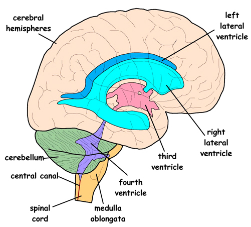
The Ventricular system
The Venous sinuses
The Reciprocal Tension Membrane System

The Sacrum and Spine

Sacral Release

Spheno-Basilar release
Temporal release
Release of Tentorium Cerebelli
Still Point Induction

Professional Insurance: Students can apply for Professional Insurance if they wish to.

Osteopathy in the Cranial Field, OCF, Course
Clinical Training
Total Hours:  Osteopathy in the Cranial Field, OCF
Location: Bahia, Brazil.

Course Fees:  £1,200


1. Scientific Study Hours:         100 Hours
3. Clinical Training Hours:          50 Lessons (à 45 min.) during 7 days

Time Table
The course runs for 7 days

    Osteopathy in the Cranial Field, OCF


What Is Osteopathy In The Cranial Field


Osteopathy in the Cranial Field (also called cranial-sacral therapy) was taught by the founder of Osteopathy Dr AT Still, MD, DO and was further developed by Dr William Garner Sutherland, D.O. (1873–1954). Was developed as an extension to the osteopathic approach over the course of 50 years by Dr. Sutherland who developed the key concepts of the Cranial Approach.

The cranial concept, first put forth by W. G. Sutherland, DO and originally alluded to by A.T. Still, MD, involves the application of Dr. Still's principles of Osteopathy to the head (cranium) and to the tailbone (sacrum). It is based on The Five Components of the Primary Respiratory Mechanism.

The "cranial osteopath" is not preferential to the cranium or the sacrum. Instead he or she includes these areas in an overall evaluation and treatment plan, considering the whole body as one dynamic, integrated unit of function.

It has become a specialized technique based on the belief that the tissues surrounding the brain and spinal cord undergo a rhythmic pulsation. This “cranial rhythm” is supposed to cause subtle movements of the bones of the skull.

The Clinical Practitioner of Osteopathy in the Cranial Field or Cranial-sacral Therapy should be able to detect these rhythms and gently manipulate the bones in time with it.

The application of Treatments aim at:

1. Relaxing Nervous Tension
2. Equalizing Rhythm of the Cerebrospinal Fluid
3. Equalizing Circulation of the Blood Flow
4. Equalizing Circulation of the Lymph Flow
5. Transfer of Body Energy (heat and vibration)

The application of which are aim at making a positive impact on the Anatomical and Physiological workings of the Body.

Osteopathy in the Cranial Field

William Garner Sutherland DO (1873-1954) graduated from the American School of Osteopathy (ASO) in 1900. As a student of Dr. Andrew Still, he took the admonition to "keep digging" very seriously. As a senior student at ASO, Dr. Sutherland happened upon a disarticulated skull in the North Hall, noting the beveled artiulcatons of the spheno-squamous suture. He had an inspiration that the bony cranium was capable of respiratory motion.

"Beveled... like the gills of a fish... indicating articular mobility... for a respiratory mechanism."

This "guiding thought" was initially quite confusing, but Dr. Sutherland could not let the idea go. Throughout his years of practice, with diligent research and study, he gradually developed a revolutionary expansion of the osteopathic concept.

Dr. Sutherland's first public announcement of his new cranial ideas were presented in September 1929, before a district meeting of the Minnesota Osteopathic Association, in a paper titled "Bedside Technique" by Blunt Bones Bill.

Dr. Sutherland called his discovery "Osteopathy In The Cranial Field" (OCF), implying that he did not create anything new. He had simply applied osteopathic thinking and principles to the cranium.

With experience, research, and a deepening of his understanding Dr. Sutherlands concept of the cranial mechanism matured. At the core of this new understanding lies the concept of PRIMARY RESPIRATION.

The definition of the Primary Respiratory Mechanism (PRM):

1. Primary: it is a system that comes "first." It underlies all of life's processes and gives dynamism, form, and substance to all of anatomy and physiology.

2. Respiratory: It is the spark that gives rise to the breath, as it moves through the tissues. It is the foundation of metabolism. It has both an inhalation and exhalation phase.

3. Mechanism: It is a system composed of many parts that work together to create a whole, greater than the sum of the parts.

The beauty of Primary Respiration is the ability to experience it directly. It is not simply a sublime concept. In the hands of a skilled practitioner, one connects directly with Primary Respiration to bring about a therapeutic response. It is the guiding principle; it is the inherent intelligence within.

Five Components of The Primary Respiratory Mechanism (PRM)

The Primary Respiratory Mechanism has classically been described as consisting of fIve components. These phenomena are expressions of involuntary physicologic motion within the central nervous system and it's adjacent anatomy.

Each component of the Primary Respiratory Mechanism, however, exists only in its relationship to the function of the whole. In truth, Primary Respiration expresses itself through all of the body, and the whole of nature. It might be said that this intrinsic motion is a fundamental expresion of life itself. Like most naturally occurring phenomenon, it is only the human mind that reduces the pieces for observation and study.

Each phenonemon is briefly described below:

The Inherent Motility of the Brain and Spinal Chord

The brain and spinal cord undulate rhythmically like a jelly fish. As the brain coils and uncoils, the cavities within the brain (ventricles) and around the brain (cisterns) change shape. During the inhalation phase (flexion) the brain (and bony cranium) gets shorter and wider. During the exhalation phase (extension) the brain (and bony cranium) gets taller and narrower.

The Fluctuation of the Cerebro-Spinal Fluid (CSF)

It is well established that the cerebro-spinal fluid (surrounding the brain and spinal chord and filling the ventricles) fluctuates rhythmically. This rhythmic fluctuation can be visually observed (and pressure changes measured) during a typical spinal tap, and has been documented by numerous research studies.

A controversial idea unique to Osteopathy describes CSF movement throughout the body, passing along the spinal nerve sheaths and through extra-cranial lymphatics. Though initially controversial, this component of the osteopathic concept is now being strongly supported by recent neuro-science research.

The CSF is considered to play a very potent nutritive role for all the tissues of the body. In his later years, Dr. Sutherland was so impressed with the potency of CSF, he would refer to it as "Liquid Light."

The Dynamic Shifting of Tensions in the Dura Mater

The Meninges are the membranes that surround the brain and spinal chord, and contain the cerebro-spinal fluid. The Dura Mater is the toughest and most outer layer of the meninges. The Dura Mater also has an internal architecture comprised of 3 sickles.One runs back to front (called the falx cerebri) and separates the two cerebral hemispheres. There are two other sickles, one on each side of the falx (called the tentorium cerebelli) that also run from the back to the front, somewhat parallel to the floor (when you are standing straight). The tentorium cerebelli gets its name from being "tent" shaped. The dura covering the outside of the brain then attaches firmly to the foramen magnum and upper cervical vertebrae, surrounds the spinal cord and descends to attach to the sacrum at the 2nd sacral segment.


The "Tent" and "Falx" (brain cut away)

These dural membranes are under constant tension. They are taut. Because of the tension through them, we refer to the Dura Mater as a Reciprocal Tension Membrane (RTM). If you pull on one end of the membrane, that pull is transmitted throughout. The Dura maintains the structural integrity of the bony cranium (as it attaches into the bones). Movement of the brain, and CSF becomes translated into the membranes as a dynamic shifting of the reciprocal tension.

So when the head is traumatized, the dural membranes may become twisted and compressed. The fulcrum around which the rhythmic movement is organized... becomes deranged. Often these membranes are the primary seat of the cranial dysfunction, actively distorting the bones they support.

The Articular Mobility of the Cranial Bones

There are 22 bones in the cranium (not including the mandible or ossicles of the ear). They meet at the suture lines. These bones form in membrane and cartilage. At birth the bones are not fully formed and are in fact quite far apart from one another. As the infant is squeezed through the birth canal, the bones slide over one another and re-expand afterwards to resume their normal positions. This physiologic compressibility allows for maximum brain capacity and minimizes brain damage. All during fetal development and after birth, the brain is constantly undulating and CSF is constantly fluctuating. This movement is transmitted through the membranes out to the bones. As the bones gradually grow to approximate one another they remain in constant motion. This movement keeps the sutures patent (open). The sutures contain blood vessels, nerve fibers, and connective tissue, just like any joint. The amount of movement is very tiny, 100ths of an inch.

This movement of the cranial bones is considered controversial. Conventional thinking considers the skull to be fused. The concept of a fused skull is erroneous, dating back to a paper written in 1873, known as "The Monro-Kellie Doctrine." The research supporting this doctrine is over 100 years old and was not very precise. More recent research supports our clinical observations of cranial bone mobility. Gradually, the mobility of the cranial bones is becoming a more widely accepted concept.

It has been found in some anatomic specimens that certain cranial sutures have fused. When fusion occurs, however, it is a pathological condition. Sutural fusion occurs because the cranium has received an impact causing the bones to compress, and lose their sutural mobility. When any joint in the body becomes immobile, it will fuse. (Put your arm in a sling, and after some time you'll lose motion in your shoulder). In health, the Cranial Sutures are patent and allow for a slight amount of motion.

Another wrinkle... We consider the bones themselves to actually be a very dense fluid. They are living tissue. They are constantly being reabsorbed and recreated. They have inherent flexibilty. Traumatic influences not only compress bones together, but cause them to "stiffen" and lose their fluid nature.

The Respiratory Motion of the Sacrum Between the Illia

The sacrum is connected to the cranium via the dural membranes that surround the spinal chord. The dura between the occiput and sacrum is referred to as "the core link." Attached only at the base of the skull and the sacrum at the 2nd sacral segment, the motion of the occiput is transmitted freely to the sacrum. The relationship of the sacrum with the occiput becomes clinically relevant when the cause of headaches may be due to a fall on the tailbone many years previously.

Dr. Sutherland was the first to discover and reason the activity of these well known anatomic structures. This model of physiologic activity provides an introduction to the "therapeutic process," and an introduction to the experience of Primary Respiration.

A skilled Osteopath is able to perceive and work with any part of this physiologic mechanism. As incredible as this may seem, we feel the bones move, the membranes pull, the fluids fluctuate, and even the brain undulate. During treatment each Osteopath may find their focus to settle upon a different aspect of this PRM. Some Osteopaths naturally engage or are more comfortable with the bones, some with membranes, some with fluid.

As I sit with my hands upon the patient, I learn how trauma has established itself in the whole person. Then as needed, I move into bone, membrane, fluid, or potency...
      Disclaimer

Please Note

The Osteopathy in the Cranial Field Course, does not, nor it is intended or qualify you as a "Doctor in Osteopathy".

In England Under Section 32(1) of the Osteopaths Act 1993 it is an offence to infer or imply that you are an Osteopath if you have not carried out the relevant training, and the following designations or titles are prohibited to use: "Osteopath, Osteopathic Practitioner, Osteopathic Physician, Osteopathist, Osteotherapist, or any other kind of Osteopath".

Successful Qualified Students after the completion of the program course in Osteopathic Manipulation, when describing themselves, either in written or verbal form, must state that they are trained, certified or qualified in Osteopathic Manipulation Techniques.


Example:

Paul Health, LMT®, CST
Lymphatic Manipulation Therapy
Osteopathy in the Cranial Field

Or

Paul Health, LMT®, CST
Lymphatic Manipulation Therapy
Cranial Sacral Therapy


Disclaimer

Graduates from Volksmed School of Health and Pharmacology having obtained the School Private Diploma of “Osteopathy in the Cranial Field” or similar, should take care not to imply that they hold a general “Osteopathic” qualification in case they do not.

The title of “Osteopathy in the Cranial Field” aren't Osteopathic doctors, and aren't registered with the General Osteopathic Council.

Thus the following information should be written in every letter and in every individual web page, this is in every page of the Practitioner webpage. This to avoid at all cost any confusion.

Please Note: Osteopathy in the Cranial Field Practitioner aren't Osteopathic doctors, and aren't registered with the General Osteopathic Council.


“Osteopathy Teaches, and has for its object the keeping and maintaining a complete circuit of the forces of the Motor, Sensory and Sympathetic Nerves, to and from the Brain and all the organs, tissues, blood and other vessels, the bowels and all parts, and the whole of the human system that pertain to nourishment, strength and growth of bone, the skin appendages and soft parts of the body.

Health is the result of the harmonious action of the system when all parts are unirritated by any cause, such as increased or diminished flow of the fluids of the arteries or veins or the nerve force, by partial or complete dislocation of bones, muscles, tissues, membranes or parts of the whole system. The object of Osteopathy is freedom of flow of all electric of other fluids, forces or substances pertaining to life
.” - Statement of Dr A.T. Still, DO upon the Diplomas of Osteopathy in 1893.

Please note: Volksmed School is not related, nor is affiliated with any other Institution teaching Osteopathy in the Cranial Field, the School if part of the Hospitallers Order of the Good News in the United Kingdom.

Contact Us


United Kingdom Registered


   Principado da Boa Nova
   Essenia
   Bahia, Brasil


  volksmedicine@gmail.com


  United Kingdom Registered

UKPRN: 10055317


School of Health & Pharmacology © VOLKSMED ® Est 2014